
How Inflammation Lowers Testosterone: Breaking the Cycle
This post contains affiliate links. As an Amazon Associate, we earn from qualifying purchases.
Inflammation lowers testosterone through three primary, documented mechanisms: pro-inflammatory cytokines like TNF-α and IL-6 directly suppress Leydig cell steroidogenesis; systemic inflammation disrupts the hypothalamic-pituitary-gonadal (HPG) axis feedback loop; and oxidative stress activates pathways like p38 MAPK that shut down key testosterone-producing enzymes. A single inflammatory insult can drop testosterone levels within hours, and chronic inflammation creates a self-reinforcing cycle that progressively dampens hormonal output.
That drop is not just a correlation, it’s a direct, causal relationship proven in both human and animal models. The body treats significant inflammation as a survival threat, and reproduction is a luxury it temporarily shelves. This isn’t about feeling a bit run down; it’s about your endocrine system receiving a chemical signal to dial back production at the source.
What follows is a breakdown of the exact biological pathways, from cytokine to cell nucleus, that explain how inflammation lowers testosterone. We’ll look at the evidence from key studies, identify the practical signs that this cycle might be affecting you, and outline a prioritized approach to reducing inflammatory load to support natural hormonal recovery.
Key Takeaways
- Acute inflammation can lower testosterone within 6 hours. Studies using endotoxin (LPS) challenges show a rapid decline in plasma testosterone, independent of other hormone changes, proving a direct attack on Leydig cell function.
- Cytokines TNF-α and IL-6 are the primary culprits. These inflammatory signaling molecules bind to receptors on Leydig cells, suppressing the expression of critical enzymes like StAR and P450scc needed to produce testosterone.
- Chronic, low-grade inflammation is a major driver of age-related low T. Conditions like obesity and metabolic syndrome create a state of constant immune system activation, which steadily erodes testicular steroidogenesis and disrupts the HPG axis over time.
- Fix the source before masking symptoms. Using supplements or stimulants to counter low-T symptoms without addressing underlying inflammation is like revving a cold engine, it might sound better for a moment but causes more wear. Bloodwork to check hsCRP is a more useful first step than any pill.
- The testes are uniquely vulnerable. Research shows inflammatory stress reduces four key steroidogenic genes in the testes, but only one in the adrenal glands, highlighting why male hormones are often the first casualty.
The Three Core Mechanisms: A Breakdown
The link isn’t mysterious. When researchers want to study how inflammation lowers testosterone, they administer lipopolysaccharide (LPS), a component of bacterial cell walls, to induce a controlled, acute inflammatory response. What happens next is a predictable cascade.
How Inflammation Directly Attacks Leydig Cells
This is the most straightforward pathway. Your immune cells, when activated, release proteins called cytokines. Two in particular, tumor necrosis factor-alpha (TNF-α) and interleukin-6 (IL-6), act as molecular saboteurs for testosterone production.
The Technical Snippet: In vitro studies exposing TM3 Leydig cells to inflammatory cytokines show TNF-α significantly decreases cell viability and testosterone production in a dose-dependent manner. In vivo, LPS treatment in mice reduces testicular gene expression of steroidogenic acute regulatory protein (StAR), cholesterol side-chain cleavage enzyme (P450scc), 3β-hydroxysteroid dehydrogenase (3β-HSD), and cytochrome P450 family 11 subfamily B member 1 (Cyp11b1), all essential for converting cholesterol into testosterone.
Think of Leydig cells as a factory assembly line. StAR is the foreman who brings the raw material (cholesterol) into the factory. P450scc, 3β-HSD, and Cyp11b1 are the specialized machines on the line. When TNF-α and IL-6 flood the facility, they first tell the foreman to stop work (diminishing StAR expression). Then they start powering down the machines. The result is a steep, rapid drop in output.
This isn’t theoretical. The 2022 mouse study in Frontiers in Endocrinology quantified it: LPS treatment caused a reduction in testicular StAR gene expression (p < 0.05), P450scc (p < 0.001), 3β-HSD (p < 0.01), and Cyp11b1 (p < 0.05). Plasma testosterone levels fell in lockstep.
Where this goes sideways: Believing you can “out-supplement” this direct suppression. If cytokines are actively blocking the enzymatic machinery, adding more cholesterol (via dietary fats) or vitamin precursors does nothing. You have to turn off the inflammatory signal first.
The Oxidative Stress and Signaling Pathway Disruption
The second mechanism is more insidious and long-term. Inflammation generates reactive oxygen species (ROS), free radicals that cause oxidative stress. This stress activates specific signaling pathways inside the Leydig cell that have one job: shut down non-essential production.
I got this wrong for years. I focused on antioxidant supplements to “mop up” free radicals, thinking that was the whole fight. The real problem is the signal the stress sends.
The key pathway is p38 MAPK. When activated by oxidative stress, p38 MAPK directly interferes with steroidogenesis. ROS signaling also increases a protein called c-Jun, which inhibits the activity of Nur77. This is a critical detail: Nur77 is an orphan nuclear receptor, a master switch that turns on the genes for those steroidogenic enzymes (StAR, P450scc, etc.). Block Nur77, and you block the cell’s ability to even make the production machinery.
The process can also be suicidal for the Leydig cells. TNF-α released by activated macrophages addresses the TNFR1 receptor on Leydig cells, triggering apoptosis (programmed cell death) and activating that same p38 MAPK pathway. Fewer cells, plus suppressed function in the survivors.
| Stress Signal | Primary Target | Result on Testosterone Production |
|---|---|---|
| Oxidative Stress (ROS) | p38 MAPK pathway | Direct inhibition of steroidogenic enzyme activity |
| ROS-mediated c-Jun upregulation | Nur77 transcription factor | Suppression of steroidogenic enzyme gene expression |
| TNF-α binding to TNFR1 | Leydig cell apoptosis & p38 MAPK | Reduced Leydig cell count & suppressed function |
The Brain’s Role: HPG Axis Disruption
Testosterone production isn’t autonomous. It’s governed by the hypothalamic-pituitary-gonadal (HPG) axis, a feedback loop between your brain and testes. The hypothalamus releases GnRH, which tells the pituitary to release LH and FSH, which then tell the testes to make testosterone and sperm. Inflammation scrambles this communication.
The same 2022 mouse study provides a clear picture. LPS treatment caused higher plasma FSH levels (p < 0.05) and higher plasma LH levels (p < 0.001). This might seem counterintuitive, shouldn’t low testosterone cause LH to rise? It does, eventually. But acute inflammation first causes a broad activation of stress-related brain nuclei, including the medial preoptic area (mPOA) where GnRH neurons are concentrated.
Common mistake: Interpreting elevated LH as a sign of “primary” testicular failure only. In an inflammatory state, it can initially indicate a disturbed HPG-axis function where brain signaling is chaotic, not a simple case of the testes not listening.
The brain perceives systemic inflammation as a major stressor. In response, it alters the pulse and pattern of GnRH secretion, leading to a dysregulated release of LH and FSH. This means the testes get erratic, poorly timed signals, further compromising efficient steroidogenesis. It’s a top-down disruption compounding the direct local attack.
The Evidence: From Human Studies to Animal Models
The mechanisms are clear in the lab. But what does the human evidence show? The data confirms the lab models and reveals the scale of the problem.
The Human Endotoxin Challenge
One of the most telling human studies involved giving healthy men of reproductive age a low-dose injection of endotoxin (LPS) to simulate acute infection. The results were stark:
- Inflammatory cytokines (IL-6, TNF-α) peaked in the blood rapidly.
- Testosterone levels showed a significant decline post-injection.
- The total reduction in plasma testosterone was significant.
- Levels returned to baseline over time.
This study, cited in the American Journal of Physiology-Endocrinology and Metabolism, is critical because it shows the effect is rapid and direct. The drop happened independently of major changes in LH or FSH, pointing squarely at impaired Leydig cell function as the immediate cause. Your body can lower its testosterone production in less time than a standard workday.
The Weight of Chronic, Low-Grade Inflammation
While acute infection models are dramatic, the larger public health issue is chronic, subclinical inflammation. This is the slow burn driven by obesity, poor diet, sedentary lifestyle, and chronic stress.
A 2023 multi-ethnic study found independent inverse associations between inflammatory markers (high-sensitivity CRP and IL-6) and both total and bioavailable testosterone in men. This held true even after adjusting for age, BMI, and other confounders. The higher the baseline inflammation, the lower the testosterone.
This creates a vicious cycle, particularly with obesity. Adipose (fat) tissue, especially visceral fat, is metabolically active and secretes pro-inflammatory cytokines like IL-6. This creates a state of chronic inflammation that suppresses testosterone. Low testosterone, in turn, promotes increased body fat and reduced lean muscle mass, which further drives inflammation. It’s a textbook negative feedback loop.
The part nobody mentions: This cycle explains why some men see little benefit from testosterone-boosting supplements alone. If the underlying driver, inflammatory adipose tissue, isn’t addressed, you’re just adding a drop of water to a bucket with a hole in the bottom. The link between obesity and low testosterone is fundamentally an inflammatory story.
What You Can Do: Breaking the Cycle
Understanding the “why” is pointless without a “how to fix it.” The goal isn’t to eliminate all inflammation, that’s impossible and unhealthy, but to reduce the chronic, dysregulated immune activity that suppresses hormones.
Step 1: Measure and Assess
You can’t manage what you don’t measure. Before buying a single supplement, get objective data.
- Run the Right Bloodwork: Ask your doctor for a test that includes hsCRP (high-sensitivity C-Reactive Protein). This is a gold-standard marker for systemic, low-grade inflammation. A result above the standard reference range indicates elevated risk. Also test Total and Free Testosterone, LH, and FSH to see the hormonal picture.
- Identify Inflammatory Triggers: Keep a simple log for two weeks. Note your diet, sleep quality, stress levels, and any symptoms (joint stiffness, brain fog, digestive issues). Look for patterns. Do symptoms worsen after eating certain foods? Is sleep poor during high-stress periods?
- Check for Stealth Conditions: Common issues like poor sleep apnea, untreated type 2 diabetes or prediabetes, and gut dysbiosis are potent drivers of chronic inflammation. They often fly under the radar while silently impacting hormones.
Step 2: Reduce the Inflammatory Load
This is about removing the sources of chronic immune activation. Think of it as stopping the leaks before you try to refill the bucket.
- Prioritize Sleep: This is essential. Poor sleep elevates IL-6 and CRP directly. Aim for 7-8 hours of quality, uninterrupted sleep. This is more powerful than any supplement.
- Address Diet: Reduce processed foods, refined sugars, and industrial seed oils. Increase intake of colorful vegetables, berries, fatty fish (rich in omega-3s), and healthy fats (olive oil, avocado). Even a modest reduction in processed carbs can lower inflammatory markers within weeks.
- Manage Chronic Stress: Long-term chronic stress elevates cortisol, which is pro-inflammatory and directly antagonistic to testosterone. Daily practices like walking, meditation, or even deep breathing can lower inflammatory cytokine production.
- Move Your Body: Regular exercise is a potent anti-inflammatory. However, balance is key. Chronic excessive cardio without recovery can increase inflammation. A mix of resistance training and moderate cardio is ideal.
Step 3: Consider Targeted Support
Once the foundational lifestyle factors are being addressed, certain anti-inflammatory supplements can provide additional support. They are assistants, not substitutes.
I learned this the hard way. Stacking five new supplements at once taught me nothing about what actually worked and likely stressed my system more. Change one thing at a time, track it, then decide.
| Supplement | Primary Anti-Inflammatory Action | Consideration for Testosterone |
|---|---|---|
| Omega-3 Fatty Acids (EPA/DHA) | Precursors to anti-inflammatory resolvins; reduce TNF-α and IL-6 production. | Foundational support. Reduces the inflammatory load that suppresses Leydig cells. |
| Curcumin (from Turmeric) | Potent inhibitor of the NF-κB inflammatory pathway. | Strong evidence for reducing systemic CRP. May help protect Leydig cells from cytokine damage. |
| Vitamin D | Modulates immune cell function; deficiency is linked to higher inflammation. | Correcting a deficiency can lower hsCRP and may improve androgen receptor sensitivity. |
| Zinc | Supports immune system regulation; deficiency can increase inflammation. | Essential for testosterone synthesis, but excessive daily intake long-term can cause imbalances. |
Start simple. A high-quality fish oil and vitamin D (if you’re deficient) are excellent first steps. Natural supplements like a well-absorbed curcumin formula can be a logical next tier. The key is to add one, monitor how you feel and your hsCRP (if re-testing), and then assess.
Why Women Get More Autoimmune Diseases
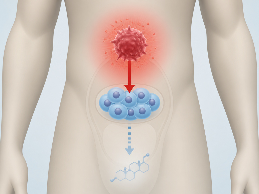
While the article focuses on how inflammation lowers testosterone in men, a broader biological pattern exists. Women have a much higher prevalence of most autoimmune diseases, which are conditions of chronic inflammation. This difference is often attributed to the complex interplay between female sex hormones and the immune system, which can create a more robust immune response. For men, the hormonal environment, particularly testosterone, is generally considered more immunosuppressive. This foundational sex difference in immune reactivity helps explain why the pathways linking inflammation to hormonal disruption, like those involving TNF-α and p38 MAPK activation in Leydig cells, are a critical concern for male health and fertility.
Can TRT Fix Inflammation-Induced Low Testosterone?
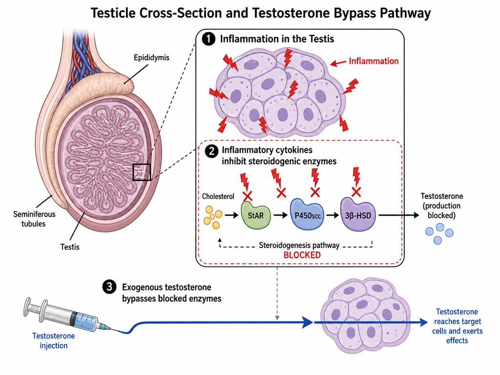
Testosterone Replacement Therapy directly addresses the hormone deficit, but it does not treat the underlying inflammatory cause. Research shows inflammation suppresses testosterone by disrupting steroidogenic enzymes like StAR, P450scc, and 3β-HSD in the testes and by activating cell stress pathways like p38 MAPK. Simply adding external testosterone may bypass the testes’ own production issues but leaves the inflammatory damage unchecked. A more complete approach involves using TRT to restore levels while concurrently working to identify and reduce the source of inflammation through diet, lifestyle, and medical management of any chronic inflammatory conditions.
Frequently Asked Questions
Can inflammation reduce testosterone?
Yes, directly and rapidly. Human studies using endotoxin challenges show testosterone can drop by about 30% within 6 hours of an acute inflammatory spike. Chronic, low-grade inflammation from conditions like obesity is a major driver of long-term, age-related decline in testosterone.
Does high testosterone cause inflammation?
No, the opposite is generally true. Testosterone has anti-inflammatory properties. It can help suppress the production of pro-inflammatory cytokines like IL-6 and TNF-α. The relationship is bidirectional: inflammation lowers testosterone, and low testosterone can permit higher levels of inflammation.
What are signs of high inflammation in the body?
Common signs include persistent fatigue, joint pain or stiffness, brain fog, skin issues like acne or eczema, digestive problems, and a tendency to gain weight, particularly around the abdomen. A blood test for hsCRP is the most objective way to confirm high systemic inflammation.
What makes testosterone go down besides inflammation?
Other primary causes include primary hypogonadism (testicular failure from injury, infection, or genetic issues), secondary hypogonadism (pituitary/hypothalamic problems), extreme chronic stress (high cortisol), significant calorie restriction, poor sleep quality, and certain medications.
Before You Go
The science is unambiguous: inflammation is a switch that turns down testosterone production. It operates through multiple, overlapping channels, direct cellular sabotage, oxidative stress signaling, and brain-axis disruption.
If you’re struggling with low energy, drive, or mood and suspect hormones, look upstream first. Chasing testosterone with stacks of supplements while ignoring a high hsCRP is the most common and frustrating mistake. The single most effective step you can take is to get that blood test. It turns an abstract feeling into a measurable number you can actually address.
Then, focus on the load. Improve sleep, manage stress, clean up your diet, and move regularly. These are the levers that control chronic inflammation. After a few months of consistent effort, retest. If inflammation markers have dropped but testosterone hasn’t recovered, then you have a clearer signal to explore more direct hormonal support with a healthcare provider. Evidence first, hype last. Start simple and stay consistent.
